Research
Evaluating optic nerve blood flow and developing new treatments and alternative diagnostic criteria for glaucoma
In addition to increased intraocular pressure (IOP), numerous studies have showed that systemic and ocular blood flow deficits are a key part of the pathogenesis of glaucoma, especially in patients with normal IOP (normal tension glaucoma: NTG). Thus, a determination of hemodynamic abnormalities in the optic nerve head (ONH) is critical to understanding the pathophysiology of NTG.
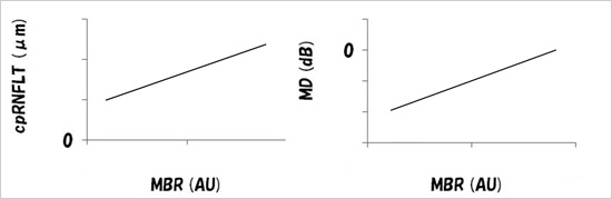
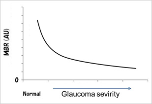
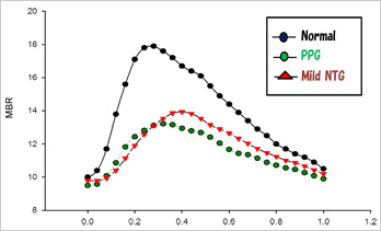
Laser speckle flowgraphy (LSFG), a non-invasive blood flow measurement technique based on the laser speckle phenomenon, provides a quantitative evaluation of ONH microcirculation that takes only a few seconds and has excellent reproducibility[1]. As we have reported, mean blur rate (MBR, an LSFG parameter representing blood flow) in the ONH has a high correlation with cpRNFLT (retinal nerve fiber layer thickness) and MD (mean deviation of the visual field measured with the Humphrey field analyzer) (Fig 1)[2]. Additionally, we found that MBR decreased with the progression of glaucoma (Fig 2), and that blood flow waveform differences (Fig 3) couldbe identified in mild NTG[3].
These results suggest that LSFG can be considered a new, non-invasive and objective instrument for the diagnosis and follow-up of glaucoma.
 |
||
| Fig 1 | ||
 |
 |
|
| Fig 2 | Fig 3 | |
Oxidative stress involvement in an animal model of glaucoma
Glaucoma is well known to be one of the world’s major causes of secondary blindness. It is thought that the ultimate cause of vision loss in this disease is retinal ganglion cell (RGC) apoptosis. A number of studies have provided evidence that oxidative stress contributes to the degeneration of RGCs in glaucoma.
Reactive oxygen species, which react with DNA, proteins and other cellular components, have been implicated in cancer, brain damage and kidney disease. NF-E2 related factor2 (Nrf2), a transcription factor activated by oxidative stress, controls hundreds of detoxifying and antioxidant genes. The function of Nrf2 in the visual system remains unclear, which prompted us to investigate the role of Nrf2 in oxidative axonal damage-induced RGC death. Seven days after optic nerve crush, the number of surviving RGCs in Nrf2 knockout mice was significantly lower than in wild-type mice (Fig 4). This suggests that Nrf2 deficiency augments NC-induced RGC death[4].
Neuroprotective strategies have been proposed and are being investigated as new goals for glaucoma therapy.
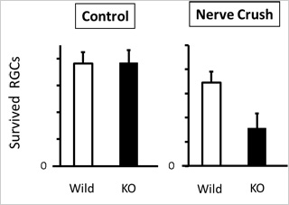
Fig 4
Worldwide research on transcriptome changes in the retina in animal models of glaucoma
Glaucoma is associated with optic nerve degeneration that results in progressive visual dysfunction. Specifically, this visual dysfunction has been reported to be the result of retinal ganglion cell (RGC) death due to axonal degeneration. However, the various pathomechanisms of glaucoma are complicated due to glaucoma’s multifactorial nature and are not yet well understood. In order to investigate the molecular mechanism of RGC death in axonal degeneration, mouse optic nerve crush models are often used[5].
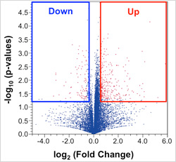
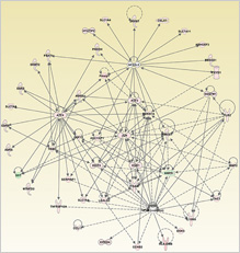
We performed RNA sequencing (RNA-seq) with a next-generation sequencer to probe global retinal transcriptome changes after optic nerve crush. We identified hundreds of differentially expressed genes (DEGs) (Fig. 5), including endoplasmic reticulum stress-related genes and antioxidative response-related genes, and revealed the global molecular network affected by axonal injury* (Fig. 6).
 |
 |
|
| Fig 5 | Fig 6 |
Masayuki Yasuda, Yuji Tanaka, Morin Ryu, Satoru Tsuda, Toru Nakazawa (2014); RNA sequence reveals mouse retinal transcriptome changes early after axonal injury. PLOS ONE (in press)
References
- Aizawa N, Yokoyama Y, Chiba N, Omodaka K, Yasuda M, Otomo T, Nakamura M, Fuse N, Nakazawa T (2011) Reproducibility of retinal circulation measurements obtained using laser speckle flowgraphy-NAVI in patients with glaucoma. Clinical ophthalmology 5:1171-1176. doi:10.2147/OPTH.S22093
- Chiba N, Omodaka K, Yokoyama Y, Aizawa N, Tsuda S, Yasuda M, Otomo T, Yokokura S, Fuse N, Nakazawa T (2011) Association between optic nerve blood flow and objective examinations in glaucoma patients with generalized enlargement disc type. Clinical ophthalmology 5:1549-1556. doi:10.2147/OPTH.S22097
- Shiga Y, Omodaka K, Kunikata H, Ryu M, Yokoyama Y, Tsuda S, Asano T, Maekawa S, Maruyama K, Nakazawa T (2013) Waveform analysis of ocular blood flow and the early detection of normal tension glaucoma. Investigative ophthalmology & visual science 54 (12):7699-7706. doi:10.1167/iovs.13-12930
- Himori N, Yamamoto K, Maruyama K, Ryu M, Taguchi K, Yamamoto M, Nakazawa T (2013) Critical role of Nrf2 in oxidative stress-induced retinal ganglion cell death. Journal of neurochemistry. doi:10.1111/jnc.12325
- Ryu M, Yasuda M, Shi D, Shanab AY, Watanabe R, Himori N, Omodaka K, Yokoyama Y, Takano J, Saido T, Nakazawa T (2012) Critical role of calpain in axonal damage-induced retinal ganglion cell death. Journal of neuroscience research 90 (4):802-815. doi:10.1002/jnr.22800
